| | ||
| STEP 1 | We will demonstrate the separation of white blood cells from whole blood for the purpose of preparing a sample for cystine analysis. 5 ml of heparinized blood was obtained from the patient a short time ago. It has been kept at room temperature no more than an hour. Transfer the blood to a centrifuge tube. If you have more than 5 ml, transfer all of it. Mark the original level of the blood (I use my thumb). (figure 1) | 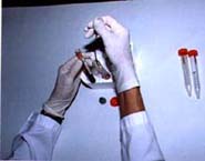 figure 1 (click on image to enlarge) |
| STEP 2 | Mix the blood with an equal volume of ACD-dextran. Do this by first adding ACD-dextran to the emptied vacutainer to a level equal to that occupied by the blood (thumb level). This will improve the yield of white cells by rinsing out the container. (figure 2) | 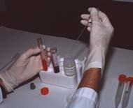 figure 2 (click on image to enlarge) |
| STEP 3 A | Add the ACD-dextran to the blood in the centrifuge tube (figure 3a) | 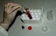 figure 3a (click on image to enlarge) |
| STEP 3 B | cap and invert to mix . . . (figure 3b) | 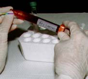 figure 3b (click on image to enlarge) |
| STEP 3 C | . . . and set the tube in an ice bath for 30 to 45 minutes. (figure 3c) | 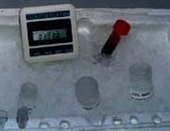 |
| STEP 4 | Leave the blood / dextran mixture on ice for 30 to 45 minutes. During that time, most of the red blood cells will settle to the bottom of the tube. At the end of that time the tube will look like this: red cells at the bottom, plasma and white cells on the top. Don't worry if the top phase looks pink. There may have been some hemolysis during the blood draw. (figure 4) | 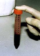 figure 4 (click on image to enlarge) |
| | ||
| STEP 5 | Transfer the top portion to a clean centrifuge tube. Avoid taking red blood cells even if some of the plasma is left. If any clots of red cells accidentally get into the sample, pull them out. (figure 5) | 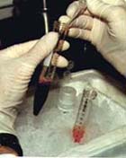 figure 5 (click on image to enlarge) |
| STEP 6 | Centrifuge the supernatant for 10 minutes to bring down the white cells. If possible, use a refrigerated centrifuge. Keeping the solutions cold helps to stablize the cystine content. For your size centrifuge use the chart in the protocol to determine the speed. (figure 6) | 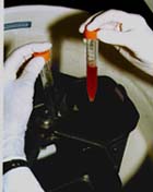
figure 6 (click on image to enlarge) |
| | ||
| Back to CYSTINE LAB TESTS & SERVICES |